
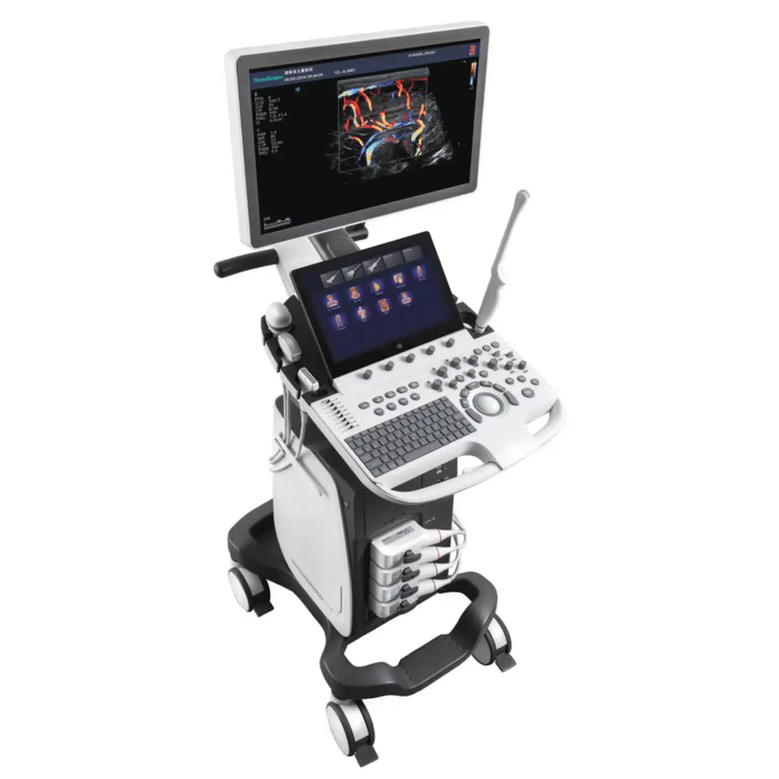
Upgraded Images with More Clarity
 |
 |
 |
 |
 |
 |
Value with Compromise, Treat with Confidence
 C-Xlasto Imaging:
With C-xlasto Imaging, P20 enables comprehensive quantitative elastic analysis. Meanwhile, C-xlasto on P20 is supported by linear, convex and transvaginal probes, to ensure good reproducibility and highly consistent quantitative elastic results.
 Contrast Imaging:
Contrast Imaging with 8 TIC curves allows doctors to assess perfusion dynamics in a wide range of clinical settings, including both the location and evaluation of lesion parts.
 S-Live:
S-Live allows for detailed visualization of subtle anatomical features, thereby enabling intuitive diagnosis with real-time 3D images and enriching patient communication.
 Pelvic Floor 4D:
Transperineal 4D pelvic floor ultrasound can provide useful clinical values in assessing the vaginal delivery impact on the female anterior compartment, judging whether the pelvic organs are prolapsed or not and the extent, determining if the pelvic muscles were torn accurately.
 Anatomic M Mode:
Anatomic M Mode helps you observe the myocardial motion at different phases by freely placing sample lines. It accurately measures the myocardial thickness and the heart size of even difficult patients and supports the myocardial function and LV wall-motion assessment.
 Tissue Doppler Imaging:
Easily Accomplish More with One Click


Reviews
There are no reviews yet.